Eye Symptoms of Stroke: What to Watch For
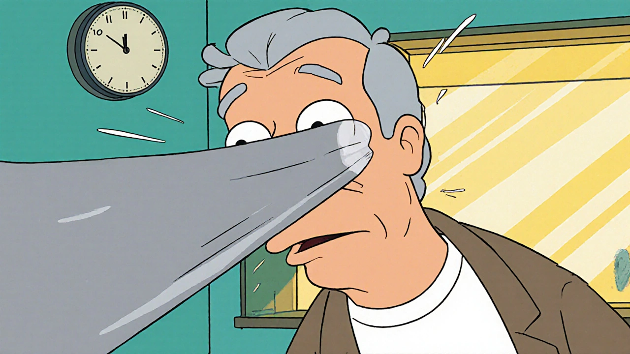
When working with eye symptoms of stroke, sudden visual changes that may signal a brain stroke. Also known as stroke‑related eye signs, it alerts you that the brain’s blood supply might be compromised. Recognizing these signs can be the difference between quick treatment and permanent damage. eye symptoms stroke are not just eye problems; they’re a window into a medical emergency.
One of the most common red flags is stroke, a sudden interruption of blood flow to the brain that can affect the visual pathways. When the optic nerve or visual cortex loses oxygen, patients may experience sudden loss of vision in one eye, double vision, or a curtain‑like shadow across their sight. Another specific sign is amaurosis fugax, brief, transient blindness in one eye caused by emboli traveling to the retinal arteries. Though it often resolves in minutes, it’s a warning that a larger stroke could follow.
Key Eye Signs That Could Mean a Stroke
Beyond amaurosis fugax, visual field loss, partial loss of peripheral or central vision is a critical clue. A sudden loss of half the visual field (hemianopia) points to damage in the brain’s occipital lobe or optic radiations. Similarly, retinal artery occlusion, a blockage of the central retinal artery that cuts off blood to the retina mimics a stroke in the eye and often heralds an imminent cerebral event. Both conditions demand immediate evaluation because the underlying cause is usually vascular.
Why do these eye signs matter? The brain and eye share the same blood vessels, so a clot headed for the brain can lodge in the retinal arteries first. Risk factors like high blood pressure, diabetes, smoking, and atrial fibrillation increase the likelihood of both stroke and eye‑related events. When you notice any of these visual changes, treat them as a possible "mini‑stroke" and act fast.
Time is brain, and time is retina. If you or someone you’re with experiences sudden vision loss, call emergency services right away. Emergency responders will prioritize rapid imaging—CT or MRI—to confirm whether a stroke is occurring. An eye exam performed by an ophthalmologist can reveal retinal artery occlusion, while a neurologist evaluates brain injury. Prompt diagnosis opens the door to treatments like intravenous thrombolysis or mechanical thrombectomy, which are most effective within the first few hours.
Treatment pathways depend on the exact cause. For an acute retinal artery occlusion, ocular massage, intra‑ocular pressure‑lowering eye drops, or hyperbaric oxygen may be tried, but the ultimate goal is to restore blood flow and prevent a full‑blown stroke. In typical ischemic strokes, antiplatelet agents, anticoagulants for atrial fibrillation, and aggressive blood‑pressure control are standard. Rehabilitation specialists also work on visual therapy to help patients adapt to any lasting field defects.
Recovery doesn’t stop at the hospital door. Visual rehabilitation can include prism glasses, visual scanning training, and occupational therapy to regain daily function. Monitoring for recurrent events is crucial; follow‑up imaging and blood‑test panels help track the health of blood vessels. Lifestyle changes—regular exercise, a low‑salt diet, quitting smoking—reduce the chance of another episode.
Below you’ll find a curated set of articles that dive deeper into each of these topics. From medication tolerance to heat safety for diuretic users, the collection covers a wide range of health issues that intersect with stroke risk and eye health. Explore the posts to get practical tips, detailed explanations, and the latest guidance on managing eye‑related stroke symptoms and related medical concerns.


